PLLA Nodule Diagnosis and Management: 2026 Consensus Guidelines for Inflammatory & Non-Inflammatory Lesions
راهنمای تخصصی تشخیص افتراقی و درمان ندول PLLA معیارهای بالینی، سونوگرافی، پیشگیری، تخریب مکانیکی، 5-FU و کورتیکواستروئید.
Introduction: Why do PLLA nodules remain a clinical challenge?
PLLA nodule formation remains one of the most challenging complications of this treatment, as its definition, nomenclature, and management are inconsistent in clinical practice. Poly-L-lactic acid (PLLA-SCA; brand name Sculptra) is the oldest and most widely used injectable biostimulator, approved in Europe since 1999 and in the United States since 2004, and is utilized for improving skin quality, restoring facial and body volume, and treating HIV-associated lipoatrophy.
In older reports, the rate of nodule formation was cited between 6% and 44%. More recent reports show a range of 0.004% to 0.4%, although these figures are derived from different study designs and denominators and should be interpreted with caution. The reduction in cases has been associated with increased dilution volumes and refined injection techniques. The frequency of inflammatory nodules (commonly referred to as “granulomas”) has been reported as 0.01% to 0.1%.
Nevertheless, the terms “nodule” and “granuloma” have been used interchangeably for years. Histology is theoretically the gold standard for diagnosis, but facial biopsy is highly invasive. Consequently, many clinicians cannot be certain whether a lesion is a product accumulation resulting from technique or a delayed host immune response. This article, adapted from a consensus guideline published in the Aesthetic Surgery Journal (2026), provides a practical framework for identifying, preventing, and managing these lesions.
Dual Clinical Definition and Classification
The consensus panel proposes using a clinical classification based on the inflammatory status of the lesion instead of histological terms, simplifying diagnosis and management in routine practice.
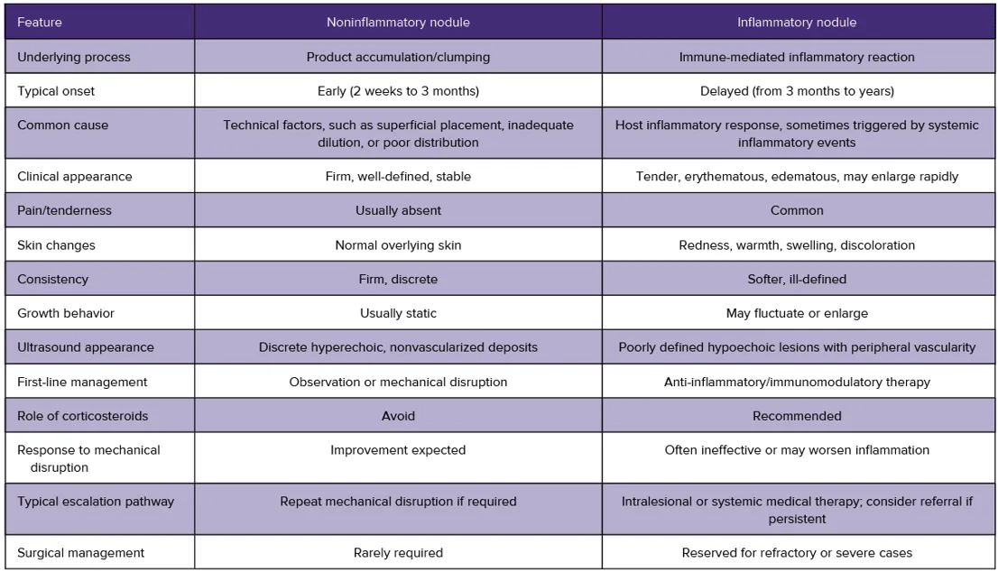
• Non-inflammatory nodule (product-related): A lesion consistent with product accumulation; typically early-onset, firm, well-demarcated, asymptomatic, and “cold” with no signs of active inflammation.
• Inflammatory nodule (host immune reaction): Formerly termed “granuloma.” Histologically, it features dense inflammatory infiltration accompanied by multinucleated giant cells. Clinically, it is “hot” and presents with tenderness to palpation, erythema, edema, and progressive enlargement.
The panel emphasizes that “granuloma” is a histological diagnosis and should only be used when confirmed by biopsy. This classification is a practical clinical framework rather than a validated pathologic classification. Furthermore, biologically, product accumulation and inflammatory reactions likely lie on a continuum with clinical overlap; therefore, classification must always be interpreted in conjunction with time of onset, imaging, and the overall clinical picture.
Diagnostic Tools: Clinical and Ultrasound Features
Ultrasound is the most practical adjunctive tool for differentiating inflammatory from non-inflammatory nodules without the need for invasive investigation. Non-inflammatory nodules appear as hyperechoic, avascular deposits, whereas inflammatory nodules present as irregular hypoechoic areas with peripheral vascularity on Doppler. Histology remains the gold standard, but performing it on facial lesions is rarely practical or justifiable, and patient consent may be difficult to obtain.
Four bedside clinical features play a key role:
Pain or Tenderness: Typically absent in non-inflammatory nodules; present in inflammatory nodules.
Overlying Skin Changes: The skin over a non-inflammatory nodule is normal; erythema, tenderness, and edema are observed in inflammatory nodules.
Consistency and Margins: Non-inflammatory nodules are firm with well-defined margins; inflammatory lesions are softer and more diffuse.
Growth Behavior: Non-inflammatory nodules typically remain stable after appearance; inflammatory nodules may appear suddenly or enlarge progressively.
The Role of Onset Time in Diagnosis
Onset time is a useful but non-definitive indicator and must be interpreted alongside clinical and ultrasound findings rather than in isolation. A practical threshold of approximately 3 months serves as a helpful guide to distinguish early lesions (likely non-inflammatory) from late-onset lesions (likely inflammatory), although it is not absolute and overlap exists.
• Early-onset nodules (2 weeks to 3 months): These are predominantly product accumulations without inflammatory reactions and are frequently related to technical factors such as dilution, injection site, or product distribution. These lesions become more prominent gradually as neocollagenesis progresses. If clinically confirmed, the initial focus is on mechanical disruption.
• Late-onset nodules (more than 3 months, up to 6 to 24 months or longer): These are predominantly inflammatory responses to foreign material that is not phagocytosed and may appear rapidly once the inflammatory response is triggered. If consistent with the clinical picture, anti-inflammatory or immunomodulatory treatment is appropriate.
Inflammatory reactions may occur months or years post-injection following a systemic immune-activating event. Specific evidence for PLLA is limited, but the panel observed that acute infections, dental procedures, vaccination, autoimmune diseases, and postpartum immune shifts can precede late-onset inflammatory nodules; this pattern is consistent with the literature regarding delayed-onset filler reactions. Therefore, when evaluating a late-onset nodule, take a careful history of recent events of this nature.

Prevention of PLLA Nodules: Minimum Standards of Care
Prevention is largely technique-dependent, and non-inflammatory nodules can be largely mitigated by modifying preparation and injection techniques.
• Dilution Volume: For facial treatments, a total volume of approximately 9 to 10 mL (8 mL of sterile water for injection plus 1 to 2 mL of lidocaine) is recommended. Lower volumes have been associated with an increased risk of nodules, and the panel considers adequate dilution to be one of the most important modifiable factors.
• Hydration Time: With sufficient mechanical agitation, hydration is virtually immediate, showing no significant differences in particle distribution or viscosity between immediate and delayed use. Immediate use following reconstitution and thorough mixing is acceptable, and prolonged hydration is unnecessary.
• Injection Depth: According to the Instructions for Use (IFU), PLLA must be placed in the subdermal or subcutaneous plane. Superficial or intradermal injection has a consistent association with early nodules and papules resulting from product accumulation. Some clinicians utilize supraperiostalar injections in specific anatomical areas; the guiding principle is to avoid superficial deposition and ensure even distribution within the appropriate tissue plane.
• High-Risk Areas: In thin tissues or dynamic areas such as the forehead, periorbital, and perioral regions, the risk of visible or palpable nodules is disproportionately high, and caution is warranted. Outside the face, the neck and hands are recognized as high-risk zones.
• Post-Treatment Massage: The "5-5-5" protocol (5 minutes, 5 times a day, for 5 days) was endorsed as a simple patient instruction and is consistent with the IFU. However, the evidentiary support for this regimen is limited, and patient compliance is variable. Thorough immediate massage by the clinician at the time of treatment was considered more critical, with patient self-massage serving a supportive role.
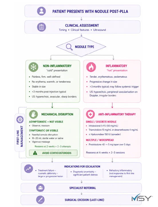
Management of Non-Inflammatory Nodules
An early non-inflammatory nodule is a product accumulation and should be treated with mechanical disruption rather than anti-inflammatory therapy.
Observation: For an asymptomatic, invisible nodule, observation and reassurance are recommended. A palpable lesion requires no intervention as long as it does not visibly enlarge or cause significant discomfort, and unnecessary treatment should be avoided.
Mechanical Disruption (first-line if intervention is needed): The nodule is disrupted with a needle or cannula, flushed with fluid, and followed by vigorous massage to disperse the PLLA particles.
Flush Fluid: Sterile water for injection and saline are both acceptable. The determining factor for success is the volume of irrigation and the degree of mechanical dispersion, not the type of solution. Because sterile water is hypo-osmolar, it may theoretically exert a greater dilutional effect via cell swelling or osmotic lysis; isotonic saline does not have this effect.
Volume: Small volumes are likely ineffective. A volume of approximately 10 to 20 mL is required, depending on the size and location of the nodule.
Timing: Early intervention — before collagen deposition occurs around the PLLA accumulation — is associated with better outcomes. In established lesions, the outcome is less predictable.
Repetition: A minimum interval of 2 weeks between sessions was proposed to allow tissue repair and nodule dispersion. Two to three attempts before changing strategy is reasonable, and ultrasonography should be used, when available, to measure nodule size and track its course.
Avoidance of Corticosteroids: These lesions are not immune-mediated; therefore, corticosteroids are unlikely to be effective and may cause local atrophy, which worsens the aesthetic result and makes the nodule more conspicuous. Overtreatment with corticosteroids or surgery for these lesions has been common in the past, exposing patients to unnecessary atrophy and scarring.
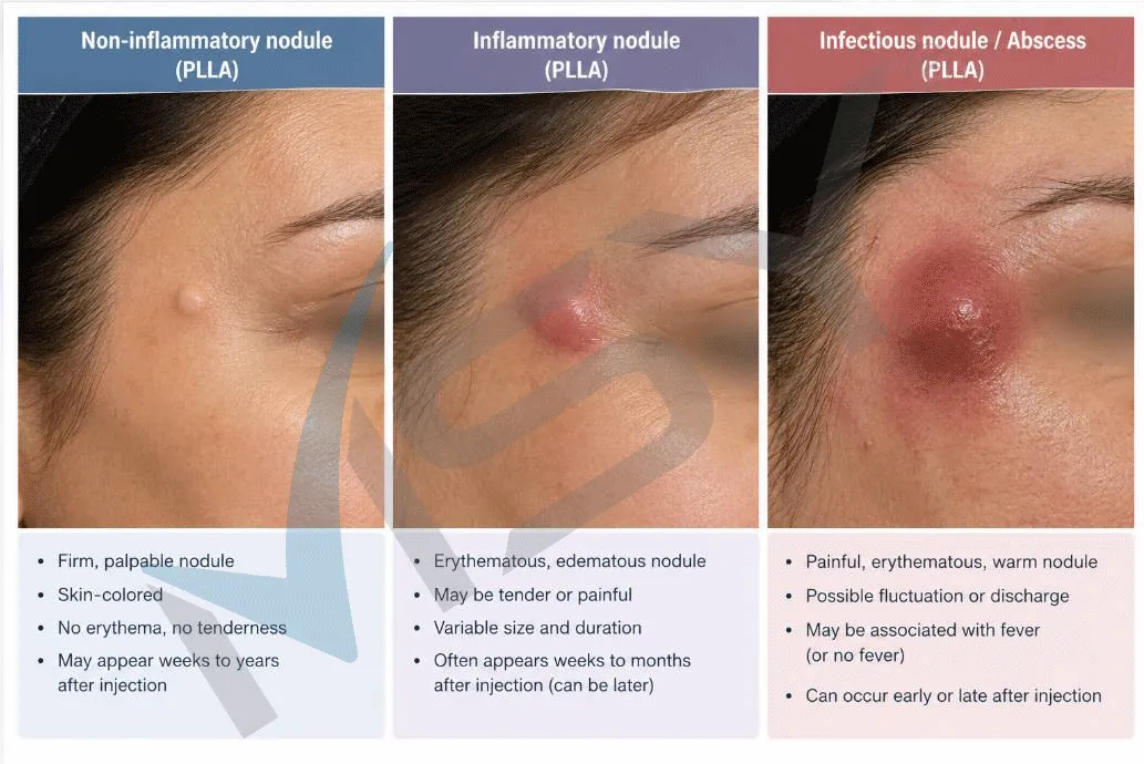
Treatment of Inflammatory PLLA Nodules
Inflammatory PLLA nodules are immune-mediated, and their therapeutic approach differs fundamentally from non-inflammatory nodules: treatment relies on anti-inflammatory and immunomodulating agents, and mechanical disruption is generally contraindicated. Based on existing evidence and expert consensus, the first-line treatment for a single inflammatory nodule is intralesional anti-inflammatory therapy.
Single or Isolated Nodule
Intralesional Corticosteroid: For example, triamcinolone (10 mg/mL) or dexamethasone (4 mg/mL); these reduce local inflammatory response, lesion size, and symptoms.
Combination Therapy with 5-Fluorouracil (5-FU): This approach is also endorsed by the panel. Off-label 5-FU—widely used in hypertrophic scars and keloids—inhibits fibroblast proliferation, targets the underlying inflammatory process, and reduces the risk of steroid-induced skin atrophy.
Common Combinations: 5-FU at a concentration of 50 mg/mL combined with triamcinolone (10 mg/mL) in ratios ranging from 1:1 to 9:1 in favor of 5-FU have been described. Adding a local anesthetic improves patient comfort.
Injection Technique: Administer into the center of the nodule using small volumes, approximately 0.1 to 0.5 mL per injection point. The 5-FU dose should remain below 1 mL, as higher volumes can cause hyperpigmentation or tissue necrosis.
Hyaluronidase: 150 units may be added as a spreading factor to enhance drug diffusion within the nodule. (Note: Hyaluronidase does not degrade PLLA).
Ultrasound Guidance: Enhances the precision of intralesional injections.
Clinical response should be assessed after a minimum of 6 weeks, and retreatment is considered only if partial improvement is observed. Two to three attempts are reasonable, and ultrasonography should be utilized where available to measure treatment response.
Multiple or Widespread Nodules
In such cases, systemic corticosteroids are more appropriate. A common regimen is oral prednisolone 40 mg with a tapered reduction to 5 mg over 5 days.
Dosages and combinations are cited from the consensus text; the final decision regarding drugs, dosing, and contraindications remains with the attending physician.
Referral, Treatment Escalation, and the Role of Infection
Treatment escalation should be based on response to first-line therapy at appropriate intervals: non-inflammatory nodules are reviewed after >2 weeks, and inflammatory lesions after at least 6 weeks. Treatment failure is defined as a persistent nodule with little to no improvement after 2 to 3 sessions of first-line therapy.
Indications for Specialist Referral:
Persistence of the lesion despite repeated first-line therapy
Symptomatic nodule
Visible deformity or significant aesthetic impairment
Large or progressively enlarging nodule
Marked patient discomfort
Diagnostic uncertainty
Referral can also be made for further evaluation, including ultrasound. Surgery is the last resort and is reserved for refractory or severe cases; excision may be definitive, but carries the risk of scarring and contour irregularities.
Infection and Antibiotics: The consensus panel does not consider delayed infection to be a major factor in typical PLLA nodules in routine practice. Acute infection resulting from a compromised skin barrier usually occurs within the first few days. While biofilms are a recognized concern for cohesive gel fillers such as hyaluronic acid, PLLA is delivered as discrete biodegradable microparticles lacking a cohesive matrix to support bacterial colonization. Therefore, the panel advises against routine empirical antibiotics for delayed inflammatory nodules, warning that misdiagnosing infection leads to polypharmacy and futile courses of antibiotics.
Informed Consent & Limitations
Informed Consent Process: The consent process must specifically cover four key points:
The small but real risk of both non-inflammatory and inflammatory nodules,
The critical importance of post-treatment massage and patient compliance,
The typically staged and conservative management approach, which may take several sessions over weeks to months, and
The rare necessity of surgical intervention in refractory cases. Some nodules may remain refractory despite proper treatment, and complete resolution cannot be guaranteed.
Limitations: A lack of comparative evidence, underreporting of adverse events, and the difficulty of histological confirmation are primary limitations. Ultrasound is not a substitute for histology. Access to ultrasound, medications such as 5-FU, and referral pathways vary globally, and localization/adaptation may be necessary.
Conclusion and Article Download
A dual clinical classification (non-inflammatory versus inflammatory), supported by ultrasonography, simplifies the management pathway for PLLA nodules and prevents unnecessary interventions. Prevention through adequate dilution, proper injection planes, and effective massage remains the cornerstone of risk reduction. Manage non-inflammatory nodules with mechanical disruption and inflammatory nodules with anti-inflammatory or immunomodulating therapy, and refer patients in a timely manner in cases of treatment failure or diagnostic uncertainty.
You can download the original article here:
https://msypharmed.com/storage/library/2026/10/result (1).pdf
References
1. Rowland-Warmann MJ, Barrera P, Clague M, et al. Consensus recommendations on the diagnosis and management of noninflammatory and inflammatory nodules secondary to poly-L-lactic acid injections. Aesthet Surg J. 2026. doi:10.1093/asj/sjag189
2. Vleggaar D, Fitzgerald R, Lorenc ZP. Understanding, avoiding and treating potential adverse events following the use of injectable poly-L-lactic acid for facial and nonfacial volumisation. J Drugs Dermatol. 2014;13:S35-S39.
3. Palm M, Mayoral F, Rajani A, et al. Chart review presenting safety of injectable PLLA used with alternative reconstitution volume for facial treatments. J Drugs Dermatol. 2021;20:118-122. doi:10.36849/JDD.5631
4. Sigrist R, Quezada N, Villaseca M, Wortsman X. Poly-L-lactic acid palpable nodules: ultrasound role in the discrimination of accumulation of product versus granulomatous reactions. Dermatol Surg. 2025;51:1179-1181. doi:10.1097/DSS.0000000000004735
5. Li A, Long A, Fang R, Mao X, Sun Q. High-frequency ultrasound for long-term safety assessment of poly-L-lactic acid facial filler. Dermatol Surg. 2022;48:1071-1075. doi:10.1097/DSS.0000000000003548
6. Modarressi A, Nizet C, Lombardi T. Granulomas and nongranulomatous nodules after filler injection: different complications require different treatments. J Plast Reconstr Aesthet Surg. 2020;73:2010-2015. doi:10.1016/j.bjps.2020.08.012
7. Lemperle G, Gauthier-Hazan N, Wolters M, et al. Foreign body granulomas after all injectable dermal fillers: part 1. Possible causes. Plast Reconstr Surg. 2009;123:1842-1863. doi:10.1097/PRS.0b013e31818236d7
8. Baumann K, Alm J, Norberg M, Ejehorn M. Immediate use after reconstitution of a biostimulatory poly-L-lactic injectable implant. J Drugs Dermatol. 2020;19:1199-1203. doi:10.36849/JDD.2020.5228
9. Bravo BSF, Carvalho RM. Safety in immediate reconstitution of poly-L-lactic acid for facial biostimulation treatment. J Cosmet Dermatol. 2021;20:1435-1438. doi:10.1111/jocd.13597
10. Narins RS. Minimising adverse events associated with poly-L-lactic acid injection. Dermatol Surg. 2008;34:S100-S104. doi:10.1111/j.1524-4725.2008.34250.x
11. Menon TSK, Dileep JE, Kuruvila S, et al. Efficacy and safety of intralesional triple combination versus intralesional triamcinolone acetonide for the treatment of keloids: a randomised controlled trial. Indian J Dermatol Venereol Leprol. 2025;91:425-431. doi:10.25259/IJDVL_1263_2024